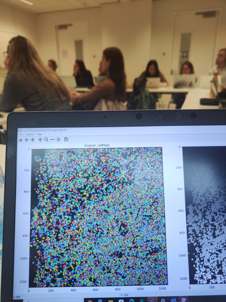
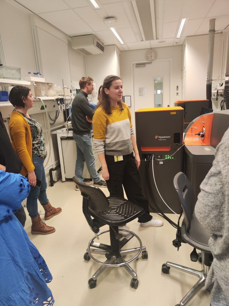
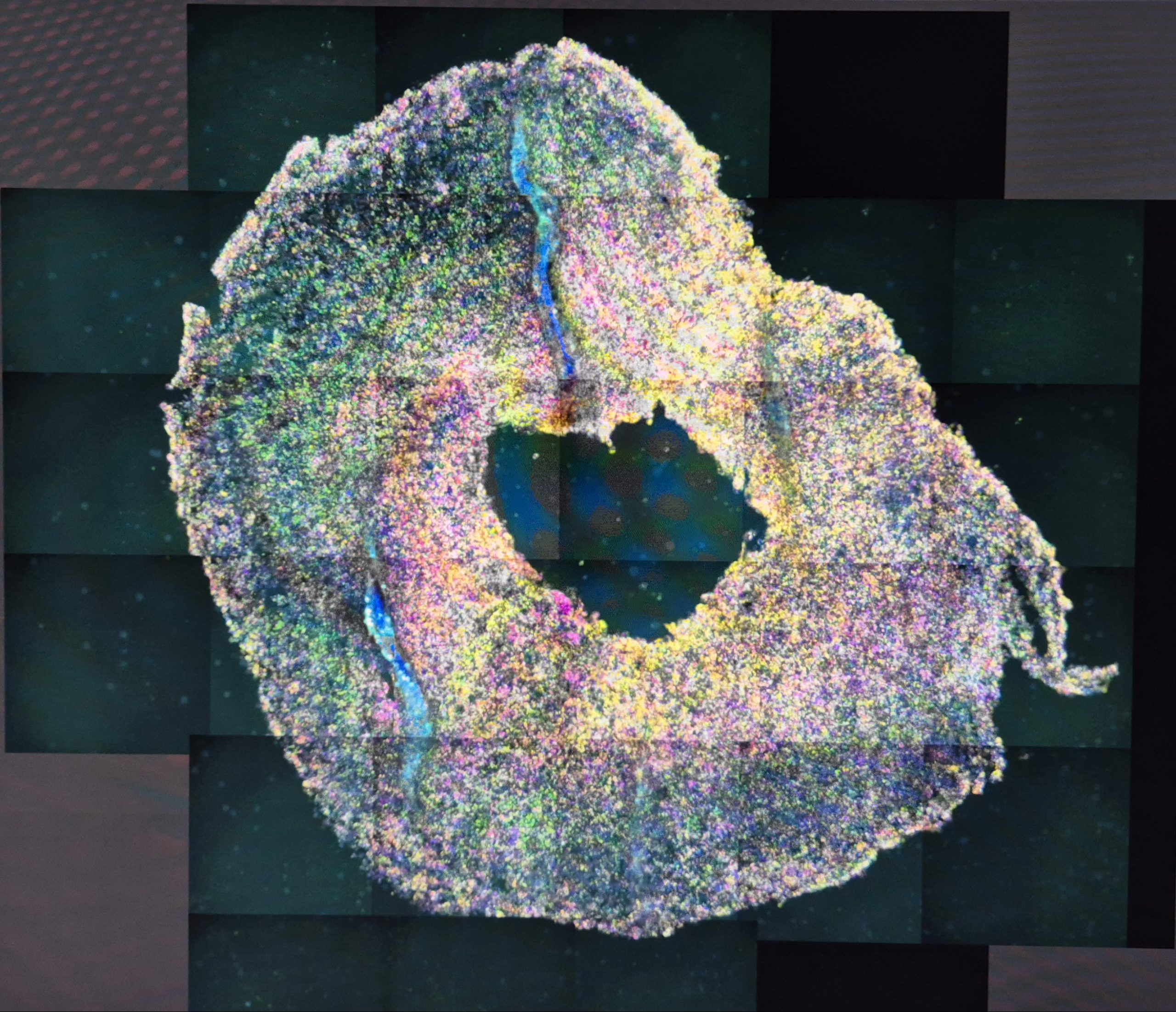
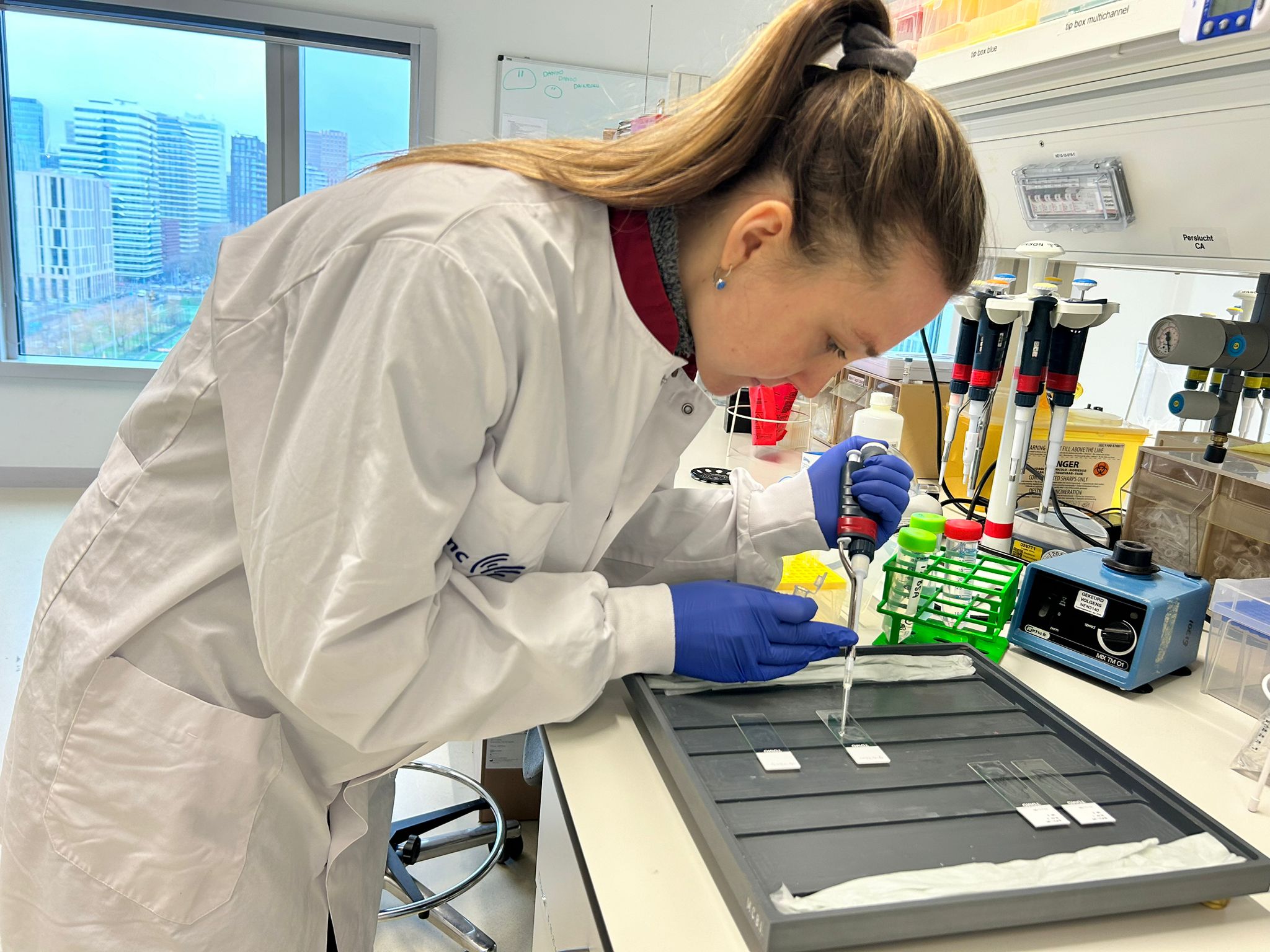
The novel technology of Imaging Mass Cytometry (IMC) enables detailed and contextual characterisation of protein expression. IMC is a mass spectrometry-based technique that allows for the immunostaining of up to 40 markers in a single tissue section. Tissue sections of primary tumours or sectioned tissue microarrays of 3D tumour models can be stained with a cocktail of antibodies conjugated to rare metal isotopes. Dr. Van Maldegem from VUmc is one of the pioneers in using and developing methods for this technology and has all corresponding infrastructure to perform IMC. Pioneering in optimisation of antibody panels to simultaneously detect tumour, immune cells, activation markers and stroma in mouse models for non-small cell lung cancer and now assembling antibody panels for the use on human tissues. Recent research focus is in overseeing the development of an automated and scalable image segmentation pipeline with which the highly multiplex IMC datasets can be analysed. Running the images through this pipeline generates very rich single cell datasets containing both phenotypic and spatial information that can be further analysed to reveal patterns in the TME. In particular, these spatial patterns of cellular neighbourhoods can only be observed using highly multiplex imaging techniques such as IMC, and are the key to distilling the regulatory networks between cells in the tissues.
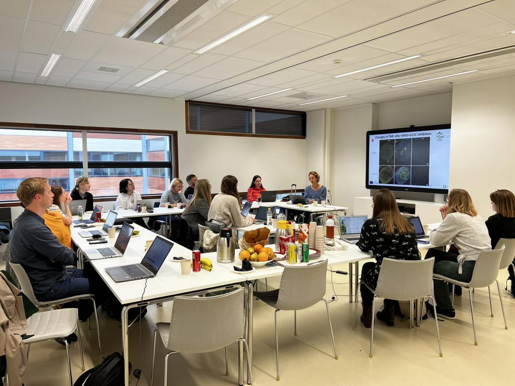
Dr. Van Maldegem, PhD Sofie Koomen and their team have organized IMC workshop for CutCancer project members. IMC technology was apply to GB samples and during the workshop we were introduced with the IMC analysis pipeline that includes panel design, segmentation, data pre-processing, sequencing segmentation, clustering, cell annotation and spatial analysis. Hands on workshop was combined with lectures from Dr. Van Maldegem, Sofie Koomen and Marieke E. Ijsselsteijn who also presented a novel data pre-propcessing tool called PENGUIN, used to analyse multiplexed spatial proteomics data.
