During 2024 EEMGS and ICAW congress in Rovinj, Croatia, researchers from National Institute of Biology presented their CutCancer related work as a posters.
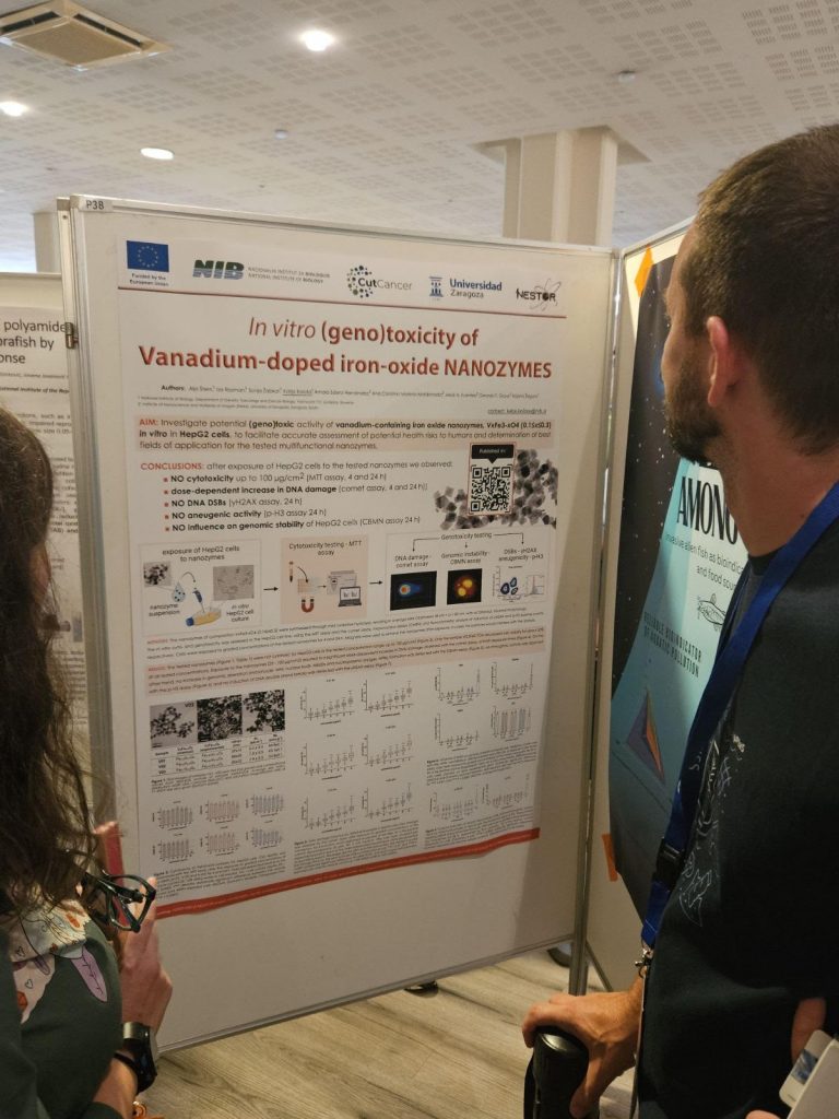
KOLOŠA, Katja, ŠTERN, Alja, ŽABKAR, Sonja, ROZMAN, Iza, ŽEGURA, Bojana, et al., EEMGS Annual Meeting and ICAW meeting, 23.-27 Sep. 2024, Rovinj. Genotoxicity assessment of Vanadium-doped iron-oxide nanozymes in HepG2 cells. Arhiv za higijenu rada i toksikologiju. [Print ed.]. 2024, vol. 75, suppl. 1, p. 145. ISSN 0004-1254.
Nanozymes, defined as nanomaterials with enzyme-like properties, pose the potential to overcome the intrinsic limitations of natural enzymes. They are characterised by being inexpensive, highly active and stable, with size-composition-dependent activity, and having a large surface area for further modification and bioconjugation. As such, they are promising candidates for use in various industries such as food processing and medicine. Vanadium ferrite (VFe2 O4 ) has attracted interest due to its catalytic and enzymatic properties. However, there is a lack of data to assess its safety, particularly regarding its potential use in biomedical and food applications. In the present study, we aimed to investigate the potential (geno)toxic activity of vanadium-containing iron oxide nanozymes VxFe3-xO4 (0.1≤x≤0.3) with octahedral, faceted morphology in average sizes D between 38 nm < D < 80 nm. In vitro cyto-/genotoxicity in the HepG2 (human hepatocellular carcinoma) cell line was assessed using the MTT assay, the comet and micronucleus assay, intracellular ROS detection and flow cytometry, respectively. Cells were exposed to graded concentrations (25–100 µg/cm2 ) of the tested nanozymes for 4 and 24 h. The results showed that the tested nanozymes were not cytotoxic to cells in the concentration range up to 100 µg/cm2, except for V0.3Fe2.7O4 , which reduced cell viability by approximately 30% at all concentrations tested. A significant dose-dependent increase in DNA damage was observed with the Comet assay at both exposure times and for all tested nanozymes. In addition, a 4-hour exposure to nanozymes led to an induction of reactive oxygen species (ROS). On the other hand, no increase in micronucleus formation or induction of DNA double-strand breaks was observed. Although further studies are required to fully clarify the safety profile of nanozymes, this study paves the way for the use of the tested nanozymes in various areas of application.
RAVNJAK, Tim, ŠTAMPAR, Martina, AUDEBERT, Marc, ŽEGURA, Bojana, EEMGS Annual Meeting and ICAW meeting, 23.-27 Sep. 2024, Rovinj. Cyclosporin A, a non-genotoxic carcinogen – its possible mechanisms of action. Arhiv za higijenu rada i toksikologiju. [Print ed.]. 2024, vol. 75, suppl. 1, p. 169. ISSN 0004-1254.
Non-genotoxic carcinogens are chemicals that can cause cancer without directly targeting DNA and altering DNA sequence. Instead of causing mutations in genetic material, these carcinogens usually interfere with cellular processes or disrupt normal regulatory mechanisms, ultimately leading to uncontrolled cell growth and tumour formation. Unlike genotoxic carcinogens, which directly damage DNA, non-genotoxic carcinogens may act through multiple mechanisms, such as epigenetic alterations, stimulation of inflammation, interfering with hormonal signalling pathways or interfering with cell division processes and migration. Here, we investigated the toxic effects of the non-genotoxic carcinogen cyclosporin A (CYC) and the negative control ampicillin trihydrate (AMP) on an in vitro 3D cell model (spheroids) developed from human hepatocellular carcinoma (HepG2) cells. The effects of CYC (0,1, 1, 10 µM) and AMP (10, 100, 1000 µM) on cell viability (MTS assay), cell cycle distribution (Hoechst 33258), cell proliferation (Ki67), DNA double-strand break (γH2AX) and mitotic cell formation (histone H3-positive events) were investigated after exposure of 3-day-old spheroids for 24 h and 96 h. The results showed that neither CYC nor AMP affected cell viability. CYC caused a moderate but insignificant increase in cell number in the G0/G1 phase after 24 h and 96 h, which was due to the accumulation of non-dividing cells (Ki67-) in the G0 phase. Conversely, AMP did not affect the cell cycle distribution. Cell proliferation decreased dose-dependently after 24 and 96 h of exposure to CYC, while AMP only slightly reduced proliferation after 24 h. AMP did not induce DNA double-strand breaks; conversely, a slight, however insignificant, increase in γH2AX-positive cells was observed after 24 h of exposure to CYC. Mitotic cell formation was not impacted, neither by CYC nor by AMP. Nongenotoxic carcinogens are an important research issue due to their complex and not fully understood potential adverse effects, which need to be further investigated.
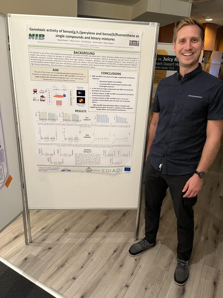
NOVAK, Matjaž, FRAS, Katarina, ŠENTJURC, Tjaša, GAJSKI, Goran, ŽEGURA, Bojana, EEMGS Annual Meeting and ICAW meeting, 23.-27 Sep. 2024, Rovinj. Genotoxic activity of benzo[g,h,i]perylene (B[ghi]P) and benzo[b]fluoranthene (B[b]F) as single compounds and binary mixtures. Arhiv za higijenu rada i toksikologiju. [Print ed.]. 2024, vol. 75, suppl. 1, p. 164. ISSN 0004-1254.
Air pollution is one of the most important environmental risk factors for health, associated with 7 million premature deaths annually. Epidemiological studies have found a strong link between air pollution and increased incidence of respiratory infections, respiratory diseases, and even cancer. Among air pollutants, polycyclic aromatic hydrocarbons (PAHs) have a high toxic potential in terms of cytotoxicity, mutagenicity, genotoxicity, and carcinogenicity. These compounds are formed during the incomplete combustion of fossil fuels, wood, coal, tobacco, and even food preparation. Consequently, PAHs can be ingested orally through contaminated food or by inhalation of polluted air. Among PAHs, benzo[g,h,i]perylene (B[ghi]P) and benzo[b]fluoranthene (B[b]F) have been identified as priority air pollutants by various environmental agencies, partly because of their frequent detection and partly because of their high toxicity. Nevertheless, information on their possible genotoxic effects is scarce. Therefore, the present study aimed to evaluate their genotoxic activity in the form of single compounds and binary mixtures (ratio 1:1) in the human hepatocellular carcinoma (HepG2) cell line, which has already been shown to be very sensitive for detecting the (geno)toxic effects of indirectly acting compounds. Cytotoxicity was determined by the MTT assay and genotoxicity by the comet and cytokinesis block micronucleus assays. The study showed that both the PAHs tested and their binary mixture can affect DNA integrity and thus pose a risk to the exposed population. Therefore, it is urgent to further evaluate their risk to a healthy population.
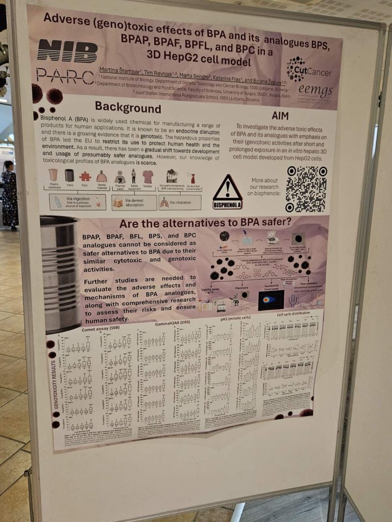
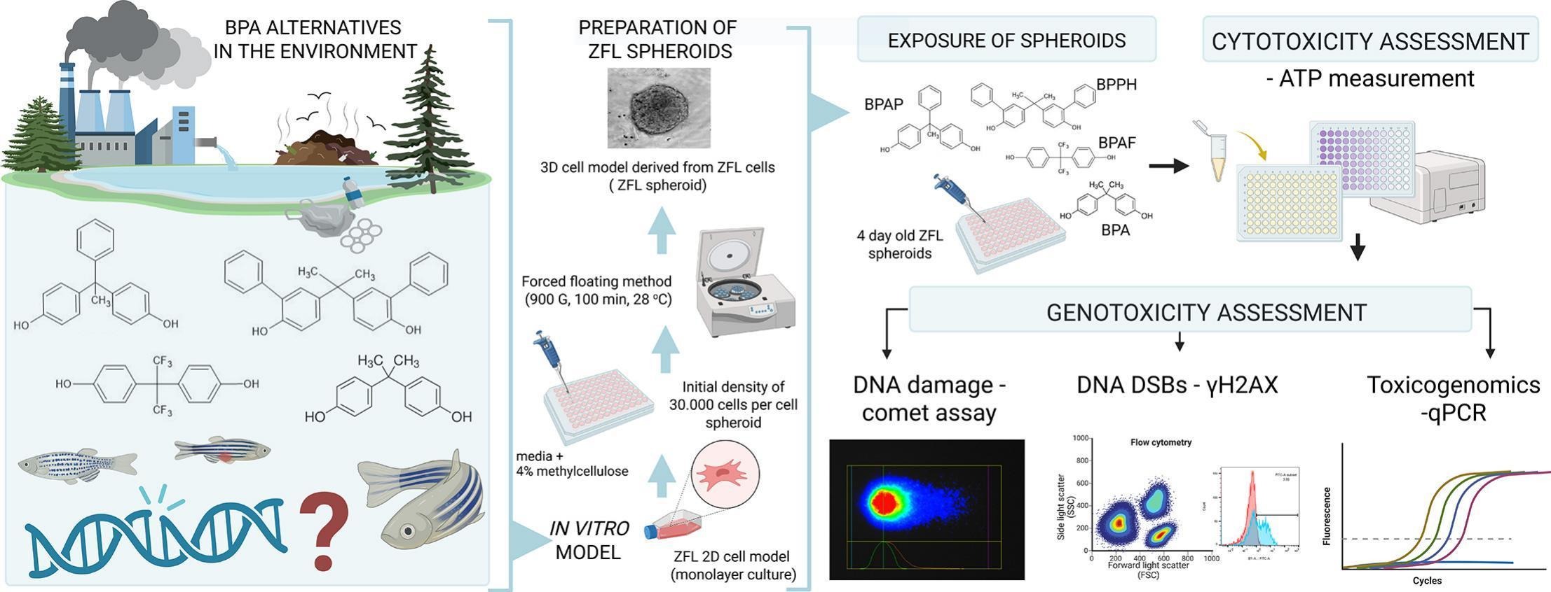
ŠTAMPAR, Martina, RAVNJAK, Tim, SENDRA, Marta, FRAS, Katarina, ŽEGURA, Bojana, EEMGS Annual Meeting and ICAW meeting, 23.-27 Sep. 2024, Rovinj. Adverse (geno)toxic effects of bisphenol A and its analogues BPS, BPAP, BPAF, BPFL, and BPC in a 3D HepG2 cell model. Arhiv za higijenu rada i toksikologiju. [Print ed.]. 2024, vol. 75, suppl. 1, p. 186. ISSN 0004-1254.
Bisphenol A (BPA) is a widely used chemical in polymer additives and epoxy resins for manufacturing a range of products for human applications. It is known to be an endocrine disruptor and there is growing evidence that it is genotoxic. The hazardous properties of BPA have raised concern and led the EU to restrict its use to protect human health and the environment. As a result, there has been a gradual shift towards using presumably safer analogues, which consequently led to an increase in their residues in the environment, however, our knowledge of their toxicological profiles is scarce. We investigated the adverse toxic effects of BPA and its analogues (BPS, BPAP, BPAF, BPFL, and BPC) with emphasis on their (cyto) genotoxic activities after short (24 h) and prolonged (96 h) exposure in in vitro hepatic three-dimensional cell model developed from human hepatocellular carcinoma (HepG2) cells. The results showed that BPFL and BPC were the most cytotoxic analogues that affected cell viability, spheroid surface area, cell proliferation, and apoptotic cell death. BPA, BPAP, and BPAF induced DNA double-strand break formation (γH2AX assay), whereas BPAF and BPC increased the percentage of p-H3-positive cells, indicating their aneugenic activity. All BPs induced DNA single-strand break formation (comet assay) (BPAP>BPFL>BPAF≈BPS>BPC≈BPA), with BPAP (≥0.1 μM) being the most effective and BPA and BPC the least effective (≥1 μM). The results showed concentration- and time-dependent effects, indicating the problem of long-term and repeated exposure of humans and animals to BPA and its analogues, in which delayed effects may occur. Overall, the present results indicate that BPAP, BPAF, BFL, BPS, and BPC cannot be considered safer alternatives to BPA in terms of their cytotoxic and genotoxic activities, and therefore further studies on their potential adverse effects and mechanisms of action are needed to adequately evaluate the risks of BPA analogues and assess their safety to humans.
ŠTERN, Alja, ŠENTJURC, Tjaša, ŽABKAR, Sonja, NOVAK, Matjaž, FILIPIČ, Metka, ŽEGURA, Bojana, EEMGS Annual Meeting and ICAW meeting, 23.-27 Sep. 2024, Rovinj. Assessment of the genotoxic and mutagenic potential of a CBD isolate and a Cannabis sativa extract in vitro. Arhiv za higijenu rada i toksikologiju. [Print ed.]. 2024, vol. 75, suppl. 1, p. 187. ISSN 0004-1254.
Cannabidiol (CBD) is a naturally occurring cannabinoid found in hemp, Cannabis sativa and C. indica plants. Unlike THC, CBD lacks psychotropic properties but exhibits various pharmacological effects. In recent years, the public perception of cannabis and CBD products has shifted, with many countries legalising their medicinal use. This shift has led to a rapid expansion of CBD products marketed as ameliorating many health problems. CBD products are used by patients as well as by healthy people who believe in the claimed benefits. While CBD’s therapeutic potential is widely reviewed, its adverse effects are less explored. The present study aimed to explore the potential genotoxic activity of a CBD isolate (CBD CP) and a Cannabis sativa extract (CBD EX). The toxic activity of the CBD samples was evaluated using the MTT assay, and the genotoxic and mutagenic activity was evaluated using the comet, micronucleus, γH2AX and p-H3 assays and the AMES test, respectively. We further performed toxicogenomic analyses of selected genes involved in xenobiotic metabolism and DNA damage response. Both CBD samples showed no mutagenic activity in the AMES test (OECD 471). They demonstrated cytotoxicity for HepG2 cells with IC50 values of approximately 26 µg/mL (4 hours) and 6–8 µg/mL (24 hours). Noncytotoxic concentrations upregulated genes encoding metabolic enzymes involved in CBD metabolism, indicating HepG2 cells’ ability to metabolise CBD. Under the applied conditions, both CBD samples were nongenotoxic, as no DNA damage or influence on genomic stability was observed in any of the applied tests (comet, micronucleus, γH2AX, and p-H3 assays), and no changes were observed in the expression of genes involved in the genotoxic stress response. The obtained results will contribute substantially to the safety evaluation of CBD-containing products and their authorisation, as well as further development in the field of hemp exploitation for medical and nutritional purposes.